Simple Bone Cyst (SBC) |Symptoms | Signs |Treatment
Also called as Unicameral Bone Cyst (UBC)


These are cavities filled with serous fluid and covered by a thin membrane in the bone.
Which age group is affected ?
- They are common in the pediatric age group of 5-20 years, boys more affected than girls.
Which bones are affected?
- Most commonly affected area is metaphysis and bone is proximal aspect of the humerus adjacent to the growth plate (nearly 60 %), followed by proximal femur, around the knee joint and rarely in the heel bone (calcaneus).
What is the cause ?
- Exact etiology is unknown
- It is suspected that it appears as a focal defect in metaphysical remodeling with blockage to interstitial fluid drainage (by Cohen).
- The cyst fluid contains prostaglandins which stimulate osteoclasts to eat away the surrounding bone, leading to the accumulation of clear yellow/straw colored fluid.
How is it Identified?
Simple Bone Cyst (SBC) |Symptoms | Signs |Treatment
- Most are asymptomatic, meaning it is diagnosed more commonly when evaluated for an episode of trauma.
- Pain primarily due to the lesion is uncommon.
- It is also common to diagnose UBC in a child when evaluating a fracture due to an episode of trauma.
- In these situations, the fracture itself acts as a stimulant to resolve and treat lesions in 8-20 % of situations with adequate immobilization depending on the bone involved.
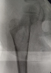
- Plain radiographs show a pure lytic lesion with a well-defined margin.
- The lesion does not usually extend beyond the original borders of the involved bone.
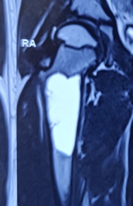
- MRI may be performed when the clinical and radiological findings allow the surgeon to suspect other causes such as Aneurysmal bone cyst, Non-ossifying fibroma or Fibrous dysplasia.
What are treatment options ?
- The natural history of these lesions are to spontaneously resolve in 33 %, increase in size during growth spurt in 33 % and reappear in 33 %.
- Usually most cysts heal by the time the patient reaches adulthood.
- It is common to note multiple episodes of fractures in individuals identified in early childhood and followed up until adulthood or maturity.
Simple Bone Cyst | Unicameral Bone Cyst
- Surgical intervention is necessary when it is situated adjacent to joints showing signs of growth (expansion), deformity, fracture, nonunion of fracture leading to weakening of the bone.
- Minimally invasive percutaneous breakage of cyst wall and 1-3 doses of steroid injections (depomedrol) under radiological guidance.
- Percutaneous curettage and infiltration of Stem cells with bone matrix.
- Curettage and filling of defect with bone graft or cement in large lesions or recurrent cases.
- Percutaneous placement of nails to drain the cyst and stabilize fracture.
- Curettage and augmentation with plate or screw and bone graft in cases of large lesion, weakened bone and proximal femur location.
- These options are individualized and none of them have been proven to be superior over other methods in clinical studies.
- In those cysts that respond to injection therapy, radiological signs of healing can be noticed anytime between 6 weeks to 6 months.
https://bonecancer.in/2020/05/18/percutaneous-decompression-curopsy/
- Results of surgical treatment are good especially when performed after the cyst has stopped growing and has become inactive.
For More Information CONTACT US

