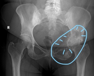
Overview & Management Options of Bone Metastases
What is Bone Metastases?
- Metastasis is considered as the ultimate step in the stage-wise progression of tumor.
- Occurrence of metastases can be regarded as either increased disease aggression or failure to respond to the treatment provided.
- Metastases can be defined as the spread of tumor cells to areas via blood vessels or lymphatic channels.
- Bone metastases comprise the majority of bone cancer burden.
- Most commonly affected are the adult and aged population.
Who are susceptible for bone metastases?
- Bone is the third most common site of metastasis after lungs and liver.
- It frequently involves individuals who have aggressive disseminated cancer disease or elderly individuals.
- 80% of patients with disseminated cancer have skeletal metastases.
- Less than 50% experience bone symptoms and less than 10% have pathological fractures.
- Two-thirds of pathological fractures in the appendicular skeleton involve the femur and the rest involve the humerus.
How does an individual with specific cancer get metastases?
- Tumor cells grow rapidly than normal cells.
- Certain factors in our body help the tumor cells produce new blood circulation thereby enhancing rapid growth and spread.
- The so called “Ligands” bind to specific receptors and alter the balance in cell signals. Bone is rich in such factors like, TGF (transforming growth factors), PDGF (platelet derived growth factors), TNF (tumor necrosis factor), parathyroid hormone-related protein (PTHrP), RANK-L (receptor activator of nuclear factor kappa B ligand) and osteoprotegerin.
- These factors are capable of supporting tumor growth which may account for the high incidence of skeletal metastases in certain types of cancers.
- RANK-L stimulates bone resorption whereas osteoprotegerin blocks resorption.
- The ratio between the two influences bone remodeling and results in ‘vicious cycle’ of osteoclast activation leading to areas of weakened bone.
What are the bones affected by metastases?
- Most metastases affect axial skeleton (includes vertebral column, pelvis, ribs and sacrum) which contain the red marrow.
- In the body, the spine is the commonest site followed by femur, humerus and pelvis.
What cancers spread to the bones?
- Carcinoma is the most common type of cancer to metastasize to the skeleton followed by myeloma and lymphoma.
- Among the carcinomas, most common are carcinomas of breast and lung (nearly 80 %), kidney, prostate and thyroid.
What are the symptoms of Bone Metastases?
- Bone metastases can result in a variety of symptoms and clinical problems which need to be either managed conservatively or tackled as a medical emergency.
- An individual with bone metastases (secondary to known cancer or sometimes, an unknown primary cancer) can present with Skeletal Related Events (SRE).
Skeletal Related Events (SRE) are as follows:
- Pain
- Pathological fractures (due to trivial trauma)
- Compression of spinal cord leading to weakness leading to disability and loss of function (Loss of bowel bladder function can also be see)
- Elevated Blood Calcium Levels
How are Bone Metastases Identified?
A detailed personal and treatment history is elicited to identify the primary cancer
Targeted investigations as follows:
- Blood Tests (Alkaline phosphatase, ESR, Lactate Dehydrogenase, Liver Function Tests, Enzyme assays specific to Cancer)
- Plain Radiographs: X-ray of the Chest and whole region involved is performed. It provides details about bone destruction or any impending pathological fracture. Lesions may be seen as sclerotic (bright and dense), lytic (hazy and dark) and sometimes mixed. There may be presence of multiple lesions along the length of the bone or spine, termed as “skip lesions”.
- MRI scan with contrast: Helps to distinguish metastases from infections in the spine.
- CT Scan: Produces excellent soft tissue and contrast resolution which shows both sclerotic and lytic lesions. Useful in spinal metastases.
- Technetium Bone Scan: Sensitive method for detection but not very specific. Useful to detect attention to areas of skeleton that require further work up.
- Whole Body PET CT Scan: To assess systemic disease load or staging. Can also be used to identify primary sources in case of a patient presenting with isolated lesion without known cause or presence of cancer.
- Image Guided Core Biopsy (may need further testing on the sample): Essential when primary is unknown or individual presents with a single suspected metastatic lesion. Not necessary when the primary cancer is identified or there are multiple lesions suspected as secondary.
- Bone lesions can be accessed by jamshidi needles, performed in a procedure room or operating theatre.
- Spinal lesions can be dealt with by needle biopsy under CT guidance or transpedicular biopsy.
- Rarely, During Emergency Surgery

For more details, please read the following different scenarios:
Scenario 1: Patient is diagnosed with Primary Cancer and Experiences Bone Pain
Scenario 2: Patient presents to hospitals/emergency with suspected fracture after trivial injury or fall (Adult/Elderly).
- Risk of Bone Metastases from Primary is evaluated- High index of clinical Suspicion if age more than 30 years, constitutional symptoms etc.
- Plain Radiograph, MRI of the involved limb is done, depending on the nature of presentation.
- Whole Body FDG PET CT scan is performed to identify any additional metastases in the body (Staging).
- If Solitary lesion-An Image Guided Core biopsy (X ray or CT or Ultrasound) is performed either by the treating surgeon or by the interventional radiologist to confirm the lesion as Bone Metastases.
- If Multiple lesions- Biopsy may not be required for confirmation.
- May need further staining (Immunohistochemistry) on biopsy samples based on pathology findings- Help in Identifying and guiding treatment.
- Discussion of the status in Tumour Board for further mode of management.
What is the significance of being diagnosed with Bone Metastases ?
- Identification of metastases and staging of the primary cancer provides an expected survival estimate with prognosis.
- Breast, prostate cancer metastases have relatively longer survival rates to lungs, thyroid, kidney, intestines due to the tendency to spread slowly.
- Functional disability based on bone/bones involved is also ascertained.
- Scoring systems such as Katagiri, Mirells, ECOG Status are used to aid treatment plans.
What is the Goal of treating Bone Metastases?
- To Relieve pain
- To Prevent fracture
- To Aid or Enhance mobility and function
- To Preserve Quality of Life
What are Treatment Options for Bone Metastases?
Bone metastases require a multidisciplinary approach and are individualised.
Treatment of individuals with bone metastases are customized and based on the following factors:
- Primary cancer histology
- Stage of primary cancer prior to metastases
- Symptoms and clinical signs
- Prognosis and anticipated survival (katagiri scoring & others)
- Bone/ Bones involved
- Extent of Metastases (Single or Multiple or Visceral)
- Functional status of the patient
- Scope for adjuvant therapy (Chemotherapy & Radiotherapy)
-
Conservative Management
Cancers which grow or metastasize rapidly have poor prognosis and are managed conservatively.
Patients whose life expectancy is less than six weeks are unlikely to benefit from major reconstructive surgery.
Various conservative strategies include:
- Analgesics (as per pain ladder)
- Chemotherapy (Conventional or Targeted based on primary Cancer)
- Radiotherapy can be provided with either “Curative or Palliative Intent” for bone metastases.
- Various modalities are Local external beam radiotherapy (single lesion), Hemi body radiotherapy (multiple lesions) and radioisotopes.
- The response of bone metastases is dependent on the primary tumor, extent of bone involvement and functional-psychosocial status of the individual.
- Individuals may experience transient or permanent pain relief within a week of radiotherapy.
- Radiotherapy is associated with complications related to gastrointestinal system and bone marrow toxicity.
- Radionuclide Therapy
- Radionuclide isotopes have also been used to treat bone metastases, especially from thyroid and prostate (strontium, radioactive iodine).
- The isotopes reach the metastases and provide palliative pain relief.
- Bisphosphonates (Ex. Zoledronic acid, Pamidronic acid, Ibandronate). Bisphosphonates are mainly used for palliative intent, targeting the osteoclastic bone resorption.
- Strengthening of a weakened bone, preventing pathological fracture
- Long term relief of bone pain.
- Hypercalcemia of malignancy
- There are also reports suggesting the delay in appearance or preventing worsening of bone metastases in patients with primary cancer.
- However, they do not play a role in improving long term survival.
- Denosumab (RANKL Inhibitor)
- Nerve Blocks
- Mobility assistive devices
-
Minimally Invasive Surgery
- Cementoplasty
- Kyphoplasty
- CT Guided Ablation (Radiofrequency or Microwave)
- Angio-Embolisation
- Surgical Interventions for bone metastases have gradually gained acceptance and popularity due to increased awareness, functional demand of the individual, improving surgical expertise, improvements in implants and prosthesis to treat such bone metastases.
- Palliative surgeries are performed for various indications such as single or dual bone metastases, compromised or weakened structure of a weight bearing bone, disabling pain secondary to metastases, extensive involvement of surrounding soft tissues, pathological fractures and impending pathological fractures.
- Single or dual bone metastasis requires a different approach in either a curative or palliative setting due to perceived prolonged survival.
- The options include Tumour Endoprosthetic reconstruction, Intercalary Endoprosthetic reconstruction, interlocking nailing of long bones and plate-cement reconstruction.
- Tumour endoprosthetic reconstruction has the advantage of allowing immediate postoperative weight bearing ambulation or mobilisation.
- Interlocking intramedullary nailing of long bones after curettage followed by cementation is performed for metastases in the long bone of the upper limb, impending pathological fracture in upper and lower limb bones and chronic pain with extensive bone metastases.
- This procedure is relatively inexpensive when compared to tumour endoprosthetic reconstruction and is a viable option in multiple bone metastases settings.
- Often, situations needing interlocking nailing or plating requires addition of radiotherapy to the affected area to provide pain palliation and also help strengthen the affected bone.
What are the advantages of Surgery in Bone Metastases?
- The ability to ambulate and perform daily activities improves self-confidence thereby attending to the psychological and social issues post metastases.
- It also reduces the risk of deep vein thrombosis, pressure sores, depression, pneumonia, constipation etc. which can happen when one is bed ridden.
-
Surgery
How are individuals with single metastases treated?
Single lesions, well documented after extensive survey and biopsy are treated with curative intent.
When found in long bones or extremities, they are customized based on functional demands of the individual.
Below are different scenarios and approach towards them:
A 55 year lady who has been operated for breast carcinoma 6 years prior has presented with pain in the hip region for 2 months.
Routine tests are performed followed by PET CT NAF/FDG and a single lesion in proximal femur is identified which is confirmed by Biopsy.
Further triple marker status is checked and prognosis estimate..
This portrays a good prognosis if metastases is approached with curative intent.
Hence, she is treated with excision of proximal femur and tumor endoprosthesis.
A tumor endoprosthesis is favored in this age group and bone involved due to the advantage of rapid return of functional status and independence.
If this individual had presented with pathological fracture, the ideal treatment would also be tumor endoprosthetic reconstruction to aid in mobilization.
Similarly, a 60 year gentleman who is being treated for lung cancer presented with sudden pain in his arm.
On investigations, a pathological fracture of humerus bone is noted.
This portrays a fair or poor prognosis with reduced long term survival.
The aim is to relieve pain and allow daily functional activities.
He is treated with extended curettage-cementing and intramedullary nailing which relieves the pain and also stabilizes the fractured bone.
A 66 year old gentleman presented with complaints of back pain.
On investigations, a single lesion was noted in the Lumbar 2 vertebrae with fracture.
Thorough workup is done to identify the primary cancer and was noted as prostate cancer.
Relevant treatment was initiated for prostate cancer, followed by aim to treat pain and subsequently mobilize.
Hence, kyphoplasty (balloon cement injection to stabilize and strengthen the vertebra) was performed.
A 48 year old lady presented with pain in her hip region for 1 month.
She was treated for breast carcinoma 3 years ago.
On investigations, a solitary lesion was identified in the acetabulum of the hip joint.
The lesion appears to be localized and not affecting the functional status, hence a surgical intervention would be morbid.
This lesion is treated by External beam Radiotherapy which relieves pain and often cures the lesion.
Alternatively, If she had presented with larger lesion and severe pain, then she would benefit from Cementoplasty or Surgery to maintain the hip integrity.
How are individuals with multiple metastases treated?
- Multiple metastases when identified portray poor prognosis and reduce the chances of long term survival.
- Supportive therapy or Palliative care is the mainstay of treatment in these individuals via multidisciplinary approach.


1 Comment